The purpose of the study was to determine relationship of abdominal fat, adipocytokine, bone mineral density, and bone turnover markers in obese male adolescents. Twenty four male adolescents (obese: 12, normal: 12) volunteered to participate in the study. Anthropometry and skeletal maturity were measured. Body composition and bone mineral density were estimated by DXA (Hologic, QDR-4500, USA). Abdominal fat with total adipose tissue (TAT), visceral adipose tissue (VAT), subcutaneous adipose tissue (SAT), and visceral adipose tissue to subcutaneous adipose tissue ratio (VSR) were estimated by computed tomography (ECLOS, HITACH, Japan). Blood samples were obtained for and analysis of adipocytokines including leptin and adiponectin. Bone turnover markers, osteocalcin (OC), bone-specific alkaline phosphatase (BALP) for bone formation markers and N-terminal telopeptide (NTx), C-terminal telopeptide (CTx) for bone resorption markers were analysed. All data were analyzed utilizing SAS 9.3 (SAS Institute, NC, USA). Independent t-test was used to evaluate the differences between obese adolescents and normal adolescents. Pearson correlation analysis was applied to figure out the relationship between abdominal fat, adipocytokines, bone mineral density, and bone turnover markers. Multiple regression analysis was used to find out the factors of abdominal fat which influence on bone mineral density. A level of significance was set at p<.05. The results of the study indicated that fat tissue (p<.001), percent body fat (p<0.001), TAT (p<.001), VAT (p<.001), and SAT (p<0.001) were significantly higher in obese adolescents than normal adolescents. However bone mineral contents were significantly higher in normal adolescents. Normal adolescents have significantly higher whole body BMD and lumber BMD than obese adolescents. Abdominal fat including VAT and SAT related negatively with whole body BMD and lumbar BMD. Leptin related negatively with BMD whereas adiponectin related positively with BMD. NTx for bone resorption marker related positively with abdominal fat. Visceral adipose tissue was a predictor for whole body BMD and lumbar BMD in explaining 46% and 32% in adolescents. In conclusion, obese male adolescents have lower whole body BMD and lumbar BMD than normal adolescents. Abdominal fat including VAT and SAT related negatively with whole body BMD and Lumbar BMD. And leptin and adiponectin were closely related with BMD. Finally, visceral adipose tissue was a predictor for whole body and lumbar BMD in adolescents.

Purpose The study examined the effects of a 12-week high intensity circuit training (HICT) on abdominal fat, physical fitness, blood lipids, and insulin resistance in middle-aged obese women. Methods Thirty obese women, aged 32-48 yrs, were recruited and randomly assigned to either HICT group (TR; n = 15) or control group (CON; n = 15). Subjects in the TR group participated in HICT of which resistance exercise and aerobic exercise were performed with a duration of 40 min/session and 3 sessions/wk for 12 weeks, whereas subjects in the CON group were asked to maintain their normal life patterns. Dependent variables included abdominal fat area, body composition, physical fitness, blood lipids profiles, and insulin resistance index. Analysis of variance with repeated measures with Bonferroni corrections was used to compare the outcomes between two groups. Results Main findings of the present study were as follows: 1) compared to the CON group, the TR group had significant reductions in overall (i.e., body mass index and percent body fat) and abdominal obesity (i.e., waist circumference, total abdominal fat area, visceral fat area, subcutaneous fat area, and visceral fat area-subcutaneous fat area ratio), 2) compared to the CON group, the TR group had significant improvements in health-related physical fitness (i.e., muscular strength, muscular endurance, muscle power, flexibility, balance, and cardiorespiratory endurance), and 3) compared to the CON group, the TR group had significant improvements in fasting lipids, glucose, insulin, and insulin resistance. Conclusions The current findings of the study suggested that HICT would be an effective exercise intervention to improve metabolic complications associated with obesity and poor physical fitness in obese middle-aged women.


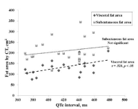
The purpose of this study was to examine the relationship between QTc interval and maximal oxygen consumption(V˙O2max) and body fat distribution in middle-aged men. Abnormal subjects (QTc interval, ≥440ms, n=10) and normal subjects(≤430ms, n=11) using QTc interval based on the Bazzet's equation were involved in the study. After overnight fasting, blood and blood pressure were measured. Abdominal fat area and regional fat compartment were measured by computed tomography(CT) and dual-energy X-ray absorptiometry(DXA), respectively. For V˙O2max, the subjects underwent a maximal graded exercise test on a cycling ergometer. Abnormal group was significantly higher in SBP, basal insulin, HOMA-IR, and leg fat compared with normal. There was a significant relationship(r=.614, p=.03) between QTc interval and V˙O2max in all subjects. Also, partial correlation analysis showed a significant relationship(r=.480, p=.032) between the QTc interval and V˙O2max. Having a QTc interval outside normal range significantly worsened risk parameters for metabolic syndrome, in particular blood pressure and insulin resistance. Moreover, QTc interval was strongly correlated with cardiorespiratory fitness in middle-aged men. This study indicates that further study will be needed to assess the exercise training effects on QTc interval.


PURPOSE This study sought to establish obesity diagnosis criteria by using the Body Volume Index (BVI) by body part extracted through 3D BodyScanner. METHODS The body fat percentage was measured using Dual Energy X-ray Absorptiometer (DEXA) for 225 participants (male = 119, female = 106), and BVI for eight body parts was measured using 3D BodyScanner. Independent t-test and Receiver Operating Characteristic (ROC) analysis were conducted. ROC analysis calculated the Area Under the Curve (AUC), and the optimal cut-point by Youden's J index. Sensitivity, Specificity, Accuracy, Balanced Classification Rate (BCR), and F1-score (harmonic mean of recall and precision) values were calculated to verify the validity of the optimal cut-point. RESULTS A statistically significant difference was observed in BVI by body part according to whether obesity was present for both men and women, and the obese group higher than the normal group. The optimal cut-point for each body part to diagnose obesity was 7.96 for shoulder, 9.79 for chest, 7.15 for upper abdominal, 7.71 for lower abdominal, 14.89 for total abdominal, 9.79 for thigh, 5.70 for calf, and 74.96 for total body volume in men. In case of women, this was 6.04 for shoulder, 9.82 for chest, 4.96 for upper abdominal, 6.23 for lower abdominal, 11.63 for total abdominal, 8.88 for thigh, 4.05 for calf, and 58.15 for total body volume, and the accuracy was 0.6~0.9. CONCLUSIONS BVI is a useful indicator for diagnosing obesity. However, this can be applicable only to Asian adults since there may be differences depending on race or age.